
Decay can be present under what would appear to be healthy tooth enamel
The photograph montage depicts a situation in which extensive decay was not diagnosed by a number of dentists prior to radiographic imaging being used.
It was not the fault of the dentists, especially since they were not initially using any dental x-rays to aid in the discovery of the decay.
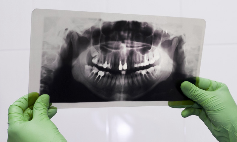
This illustration demonstrates that decay can be present under what would appear to be healthy tooth enamel and that periodic dental x-rays are necessary to reveal dental problems.
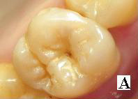
The decay was not clinically apparent to the eye and could not be detected with an explorer.
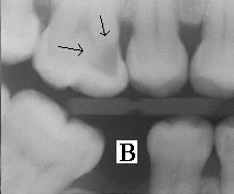
The X-Ray revealed an area of extensive region of demineralization and decay within the dentin (arrows) over one half of the tooth.
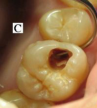
A dental burr was used to remove the enamel (what appeared to be healthy tooth enamel) overlaying the decay. A large hollow/cavity was found within the tooth. It was discovered that a hole in the side of the tooth, was large enough to allow the tip of the explorer to pass, and was contiguous with this hollow.
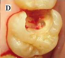
After all the decay had been removed, it was found that the decay had spread to more than half of the tooth structure and the pulp chamber had been contaminated.

Leave a Reply